


Direct Digital Radiography vs. Computed Radiography
Jump to:
- What Is Direct Digital Radiography?
- What Is Computed Radiography?
- Choosing Direct Digital or Computed Radiography System
Computed radiography (CR) and digital radiography (DR) are increasingly embraced by many industries utilizing non-destructive testing. These methods prove to be more cost-efficient compared to traditional film systems, both in acquisition and operation. In the debate of CR vs DR, the latter presents a more optimized workflow with swifter image processing.
Owing to the rapidity of DR, more images can be captured and processed within the same timeframe, resulting in a lower cost per image. DR also includes computed tomography, a technique that amalgamates multiple digital images to assemble detailed three-dimensional assessments. While this technique finds extensive use in medical applications, its utility in non-destructive testing is a recent advancement.
What Is Direct Digital Radiography?
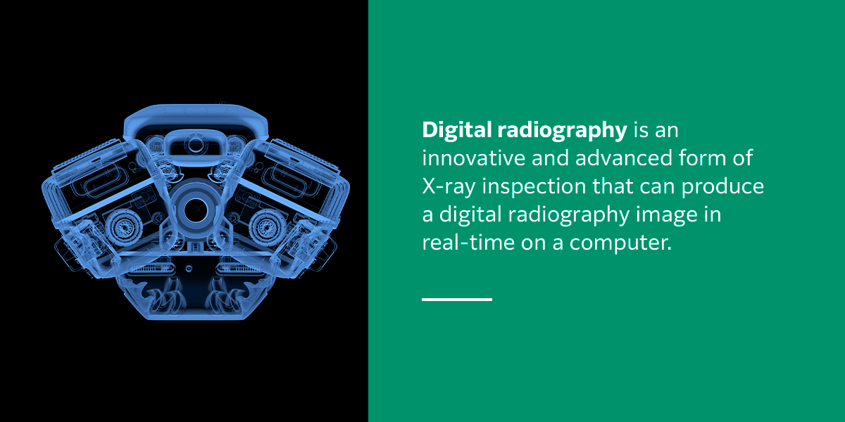
Direct digital radiography, also known as DR or DDR, is the digital registration of an image using a detector without an intermediate processing step to successfully obtain the digital signals as is needed with computed radiography. Digital radiography is an innovative and advanced form of X-ray inspection that can produce a digital radiography image in real-time on a computer. The DR process uses X-ray-sensitive plates to capture information when examining a product.
The information gained during direct digital radiography is promptly transferred to a computer without needing an intermediate cassette. The X-ray radiation is converted into an electric charge and then into a digital image via a detector sensor. Flat-panel detectors, also referred to as digital detector arrays (DDAs), can provide clear, precise, high-quality digital images. Flat-panel detectors also offer improved dynamic range and enhanced signal-to-noise ratio, providing a higher level of sensitivity for various radiographic applications.
Flat-panel detectors use both gadolinium or cesium scintillators and amorphous silicon detectors that convert X-rays to light, which transistors translate into digital information. As with CR, you can expect exceptionally high-quality images using DR. They are immediately sent to a monitor or computer in real-time. DR is often ideal for businesses or facilities with high workloads that rely on consistent and quality imaging. There are two different approaches that flat-panel detectors work on, including indirect and direct conversion.
Indirect Conversion
Indirect conversion is a technique that uses a scintillator to effectively convert X-rays to light before they are transformed to an electrical charge for readout. When X-ray photons encounter a cesium iodide scintillator, they are converted to light. The scintillator features an almost needle-like structure that is designed to minimize overall scatter at this point.
A scintillator layer can convert X-ray photons into photons of visible light. At this point, the light then reaches a low-noise photodiode array, transforming it into an electrical charge. The electrical charge created is directly influenced by the energy and number of X-ray photons that interact with the detector pixel and the density and amount of material that has effectively absorbed the X-rays. Each of these photodiodes represents a pixel, with each producing an electrical charge that will be read out digitally and then sent to the image processor.
Direct Conversion
Direct conversion is a technique that can directly convert the absorbed X-ray energy into electrical charges sized proportionally without the need for an intermediate scintillating step. Direct conversion uses a semiconductor material that creates electron-hole pairs proportional to the X-ray intensity. In direct conversion, one of the most commonly used semiconductors is amorphous selenium.
Flat-panel detectors in direction conversion processes use a photo-conductor like cadmium telluride or amorphous selenium on a multi-micro electrode plate. This helps provide the highest resolution and sharpness possible. Thin-film transistors then read the information in both detector types.
Direct Digital Radiography Applications
Direct digital radiography is an innovative technology used across many industries, including aerospace product examination, foreign object detection, casting and weld inspection and product and process development. Other applications that commonly require the use of direct digital radiography include the detection of corrosion under insulation and flow-accelerated corrosion, composites and fiber-reinforced components and various healthcare uses.
Direct digital radiography is used frequently because it offers various industries numerous benefits and advantages. With direct digital radiography, organizations can expect shorter exposure times, enhanced detail detection, real-time applications and increased portability. These advantages can provide immediate feedback and improve a facility’s overall productivity.
Direct digital radiography results are also easy to transfer between departments or even to clients electronically. DR also boasts reduced inspection time because there is no chemical processing of film required for results. With increased dynamic range, direct digital radiography can allow multiple thicknesses to be inspected at once.
Signal-to-noise (SNR) ratios are an important metric that compares the desired signal to the amount of background noise. Direct digital radiography can enhance overall SNR and linearity and uses innovative defect-recognition software and various analytic tools. Industries using direct digital radiography can also enjoy enhanced digital images and information storage.
What Is Computed Radiography?
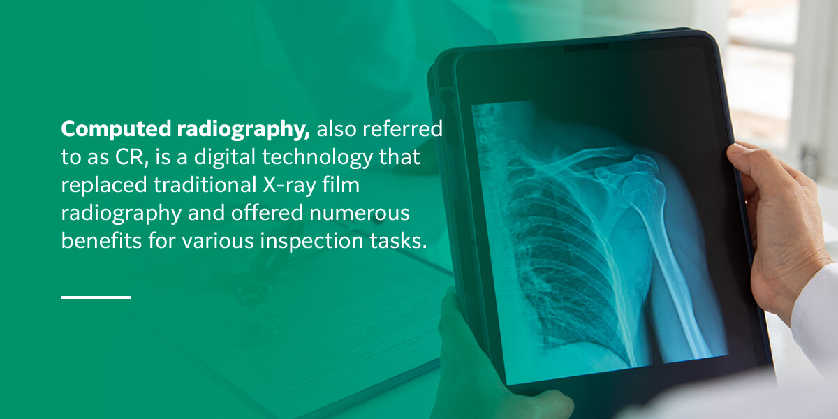
Computed radiography, also referred to as CR, is a digital technology that replaced traditional X-ray film radiography and offered numerous benefits for various inspection tasks.
Some of the main benefits of computed radiography include a reduction in processing time and exposure, ease of use, software-based assessment and reporting and simple digital archiving and information transfer. Additionally, there is no darkroom or chemicals required with computed radiography, and the imaging plates are reusable if in good condition.
Computed radiography offers a higher dynamic range when compared to traditional film, allowing more details to be clearly visible and analyzable. Additionally, computer radiography makes for a much safer working environment, streamlines various workflows and is more environmentally friendly because the entire process is chemical-free.
When an imaging plate is exposed to gamma rays or X-rays, the energy of this radiation is housed in a specialized phosphor layer. A specialized scanner can then read out the image from the plate by using a finely focused laser beam to stimulate the plate. Once stimulated, the plate can emit a blue light proportional to the amount of radiation delivered during exposure.
A photomultiplier, a sensitive analog device, detects the lights and converts the light into a digital signal via an analog-to-digital converter (ADC). The digital X-ray can be viewed and assessed on a monitor in real-time. Once an imaging plate has been viewed, it can be reset by using a high-intensity light source to erase information, allowing these imaging plates to be reused many times depending on their application and wear and tear level.
Benefits of Computed Radiography Over Film
Computed radiography is often used as a digital replacement for traditional X-ray film. Computed radiography imaging plates are used with the same techniques and radiographic inspection methods as traditional film and are available in different image qualities, which have unique required exposure times. The imaging plate type is not the only aspect that directly influences image quality with computed radiography technology.
Another essential aspect that influences image quality is the scan settings used by the scanner, specifically the overall capability of the scanner. Computed radiography’s digital magnification allows for more efficient and accurate detail viewing. Computed radiography is compatible with many traditional systems and is effective for low-volume or smaller facilities.
The digital images and information from computed radiography are securely stored and easily shared with customers or other team members. Plate exposure times with computed radiography are also significantly less than traditional film. Another important benefit of computed radiography is that it is a low-cost initial investment.
Computed Radiography System Components
A computed radiography system includes various components, including software and hardware, that accomplish several tasks, including radiographic display, radiation detection and archiving or storage. The hardware of a computed radiography system includes a laser light scanner, also known as a plate reader, imaging plates, computer and a printer.
An important aspect of computed radiography systems is the proper maintenance of the plates. Plate maintenance involves routinely cleaning plates with ethanol-soaked gauze. When cleaning computed radiography plates, it is important to be gentle with this equipment. After each exposure and read cycle, the plates will need to be erased to remove any remaining energy in the phosphor. If plates are not effectively erased after each use, they may potentially produce ghost images.
During the laser scanning processes, approximately half of the stored energy is effectively removed, making it essential that the plates are fully erased before being used again. In some systems, this process can be achieved along with image reading. It is also recommended that plates be exposed to bright white light at least once a week to erase any residual energy that may be captured as they sit unused.
In most cases, computed radiography systems also have a dedicated computer. On average, radiographs often require between one and 10 or more megabytes of space, usually stored on a network server, system hard drive or portable DVDs or CDs. Generally, DVDs are preferred because they have more storage capacity than CDs.
Choosing Direct Digital Versus Computed Radiography System
When implementing a state-of-the-art radiography system, you will want to consider if direct digital radiography or computed radiography systems may be right for your organization. To decide between these two systems, you will need to consider each of their capabilities and determine which may be right for your facility’s needs and goals.
Digital systems offer many benefits over traditional film-based systems, especially in terms of portability and sharing results. Digital systems allow for easy and simple storage of information and the ability to send this data wherever you need it in a matter of seconds. Traditional films were much more difficult to share effectively and had to be meticulously stored in humidity-controlled areas.
There are numerous factors to consider when determining which method may suit your needs. When compared to traditional film, both direct digital radiography and computed radiography require significantly less exposure. Additionally, both DR and CR systems offer high-latitude images that do not rely on various chemicals for processing.
Both DR and CR systems can offer high-quality images, depending on the overall setup, the object and quality requirements in place. To effectively compare direct digital radiography and computed radiography systems, you will want to consider the pros and cons of each system.
Pros and Cons of Direct Digital Radiography
Direct digital radiography is one of the latest and most innovative advancements within the field of radiography. Digital radiography uses a digital X-ray detector to instantly acquire images and transfer this data to a monitor for immediate viewing. Direct digital radiography is capable of both mobile and fixed use. Because of its many unique benefits, digital radiography is a more expensive option.
Direct digital radiography brings higher-quality images at a higher price and is significantly more efficient and effective than traditional systems. Because direct digital radiography has high-volume capabilities, it is generally recommended for high-volume facilities or larger businesses with a higher workload.
Some of the most beneficial aspects of digital radiography include greater dose efficiency with cesium iodide panels and high-volume capacity. Additionally, digital radiography also achieves better quality images and faster image acquisition. While these advantages are impressive and beneficial, it is also important to consider potential drawbacks associated with digital radiography.
Because of the higher price point, it is a more expensive technology to implement for a business. Additionally, direct digital radiography systems are delicate and require care and protection from mishandling and dropping. A protection cover is also generally recommended if the panel is removed from a table or bucky tray.
Pros and Cons of Computed Radiography
Computed radiography is also an innovative radiography tool that uses phosphor imaging plates to create a digital image. A computed radiography system utilizes a cassette-based system similar to along film and is often referred to as a bridge between traditional radiology methods and fully digital systems. One of the main benefits of computed radiography is that it is considered the most initially cost-effective choice for a facility.
Computed radiography functions similarly to traditional film and only requires minor changes to your workflow and facility. The overall initial investment with computed radiography is also much lower than implementing a direct digital radiography system. Although computed radiography is a beneficial system, it may not streamline processes as effectively as digital radiography. Additionally, computed radiography systems often require more maintenance.
Computed radiography cassettes also risk being mishandled, damaged or lost but are generally cheaper to replace than wireless digital radiography panels. Computed radiography may be ideal for smaller or lower-volume businesses with a low initial investment cost and has multiple sizes for increased flexibility. Additionally, CR systems are compatible with a wider range of traditional systems.
Another benefit of CR is its ability to read smaller defects. Our CR system, for example, has 25µm resolution readings, while most DDA systems offer 100µm. The CR reading has better spatial recognition for these smaller items. To achieve similar resolution, a DDA might call for geometric magnification — a process that decreases the number of smaller parts that can be scanned in one image and increases the number of images needed for larger parts. In this example, CR is more efficient than DR.
In addition to these advantages, you may also want to consider the potential drawbacks of a CR system, including its need for more intensive maintenance and potential for repairs. There is also a risk of overexposure with computed radiography systems, and they tend to take longer to view images.
Learn More About Our Digital Radiography Solutions
At Fujifilm, our mission is to use the latest, most advanced technologies to provide the highest quality products and services to contribute to the continuing advancement of science, culture, industry and technology. Our goal is to help improve the overall quality of life of people all over the world. We are proud to offer state-of-the-art products and services, including commercial technical services, data storage, optical devices, industrial materials and more.
We are also leaders in the field of non-destructive testing (NDT), which has several disciplines. One of the leading NDT disciplines is in the use of industrial radiography using ionizing radiation producing gamma rays, X-rays or neutrons. Learn more about our non-destructive testing products and services today.